Class 11 Biology · NCERT Chapter 16
~44 min readExcretory Products and Their Elimination Class 11 Notes
Exam-ready notes on excretory products and their elimination: ammonotelism, ureotelism and uricotelism, invertebrate excretory structures, the human excretory system and nephron, urine formation (GFR, JGA), the countercurrent mechanism, hormonal regulation (ADH, renin-angiotensin, ANF), micturition, accessory excretory organs and renal disorders.
What are the three processes of urine formation?
Answer-first: urine formation involves (1) glomerular filtration — ultrafiltration of blood plasma at the glomerulus (GFR ≈ 125 mL/min = 180 L/day), producing a protein-free filtrate; (2) tubular reabsorption — selective recovery of essential nutrients, electrolytes and water back into the blood, mainly in the PCT (>99% of the filtrate); and (3) tubular secretion — transfer of H⁺, K⁺ and ammonia from the blood into the filtrate, maintaining pH and ionic balance.
Modes of Excretion — Ammonotelism, Ureotelism and Uricotelism
Ammonia, urea and uric acid are the major nitrogenous wastes excreted by animals. Which one an animal excretes depends mainly on its habitat and the availability of water. Ammonia is the most toxic and requires large volumes of water for elimination; uric acid, being the least toxic and nearly insoluble, can be removed with a minimum loss of water.
Ammonotelism
The process of excreting ammonia. Many bony fishes, aquatic amphibians and aquatic insects are ammonotelic. Being readily soluble, ammonia is excreted by diffusion across body surfaces or through gill surfaces (in fishes) as ammonium ions; the kidneys play no significant role.
Ureotelism
Excretion of urea, a less toxic nitrogenous waste that needs less water. Mammals, many terrestrial amphibians and marine fishes are ureotelic. Ammonia produced by metabolism is converted into urea in the liver, released into the blood, and filtered and excreted by the kidneys. Some urea may be retained in the kidney matrix to maintain a desired osmolarity.
Uricotelism
Excretion of uric acid as a pellet or paste, with minimum loss of water. Reptiles, birds, land snails and insects are uricotelic.
- Protonephridia or flame cells — excretory structures in Platyhelminthes (e.g. Planaria), rotifers, some annelids and the cephalochordate Amphioxus; concerned primarily with ionic and fluid-volume regulation (osmoregulation).
- Nephridia — tubular excretory structures of earthworms and other annelids; remove nitrogenous wastes and maintain fluid and ionic balance.
- Malpighian tubules — excretory structures of most insects including cockroaches; help in removal of nitrogenous wastes and osmoregulation.
- Antennal glands or green glands — perform excretory function in crustaceans like prawns.
NEET favourite
MCQ Questions
1 Mark Questions
- 1Define ammonotelism with one example.
- 2Define ureotelism and name the organ where urea is formed.
- 3What are malpighian tubules? Name an organism that has them.
- 4Name the excretory structure of crustaceans like prawns.
- 5What is osmoregulation?
2 Mark Questions
- 1Why does ammonia need large volumes of water for elimination while uric acid needs the least?
- 2Distinguish between ammonotelism, ureotelism and uricotelism with one example of each.
- 3Name the four invertebrate excretory structures and their functions.
3 Mark Questions
- 1Terrestrial animals are generally either ureotelic or uricotelic, not ammonotelic. Justify, using the toxicity and solubility of the nitrogenous wastes.
- 2Survey the variety of excretory structures in the animal kingdom — flame cells, nephridia, malpighian tubules and green glands — and state the function of each.
Human Excretory System — Organs
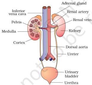
In humans, the excretory system consists of a pair of kidneys, one pair of ureters, a urinary bladder and a urethra. Kidneys are reddish-brown, bean-shaped structures situated between the levels of the last thoracic and third lumbar vertebra, close to the dorsal inner wall of the abdominal cavity.
- Each kidney of an adult human measures 10–12 cm in length, 5–7 cm in width and 2–3 cm in thickness, with an average weight of 120–170 g.
- Towards the centre of the inner concave surface is a notch called the hilum, through which the ureter, blood vessels and nerves enter.
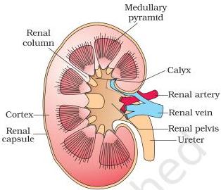
- Inner to the hilum lies a broad, funnel-shaped space called the renal pelvis, with projections called calyces.
- A pair of ureters carry urine from each kidney to the urinary bladder, which stores it until a voluntary signal causes its release through the urethra.
Exam pointer
MCQ Questions
1 Mark Questions
- 1List the organs of the human excretory system.
- 2Where are the kidneys located in the body?
- 3Give the average weight and dimensions of an adult human kidney.
- 4What is the hilum of the kidney?
- 5Define calyx and renal pelvis.
2 Mark Questions
- 1Explain the roles of the ureters, urinary bladder and urethra in urine transport and storage.
- 2Describe the hilum and renal pelvis of the kidney with their significance.
- 3Distinguish between the renal cortex and renal medulla.
3 Mark Questions
- 1Describe the position, shape, size and weight of the human kidney, mentioning the hilum and renal pelvis.
- 2Trace the path of urine from the kidney to the external urethral opening, naming each part involved.
Structure of the Kidney — Cortex, Medulla and Pyramids
The outer layer of the kidney is a tough capsule. Inside the kidney there are two zones: an outer cortex and an inner medulla. The medulla is divided into a few conical masses — the medullary pyramids — that project into the calyces. The cortex extends in between the medullary pyramids as renal columns called the Columns of Bertini.
Columns of Bertini
The portions of the renal cortex that extend between the medullary pyramids. Also called renal columns.
MCQ Questions
1 Mark Questions
- 1Name the two zones of the kidney.
- 2What are medullary pyramids?
- 3Define the Columns of Bertini.
- 4What are the projections of the renal pelvis called?
2 Mark Questions
- 1Describe the internal structure of the kidney — capsule, cortex, medulla, pyramids, pelvis and calyces.
- 2Distinguish between the cortex and the medullary pyramids of the kidney.
- 3How do the collecting ducts of the nephrons open into the renal pelvis?
3 Mark Questions
- 1Give a labelled account of the longitudinal section of the human kidney and describe the medullary organisation.
- 2Explain the relationship between the medullary pyramids, calyces and renal pelvis in draining urine.
Nephron — Structure and Types
Nephron
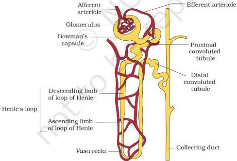
The functional unit of the kidney. Each kidney has nearly one million nephrons. A nephron has two parts: the glomerulus and the renal tubule.
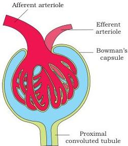
The glomerulus is a tuft of capillaries formed by the afferent arteriole — a fine branch of the renal artery. Blood from the glomerulus is carried away by an efferent arteriole. The renal tubule begins with a double-walled cup-like structure, Bowman's capsule, which encloses the glomerulus. Glomerulus together with Bowman's capsule is called the malpighian body or renal corpuscle.
- The tubule continues as a highly coiled proximal convoluted tubule (PCT).
- A hairpin-shaped loop of Henle follows, with a descending and an ascending limb.
- The ascending limb continues as the distal convoluted tubule (DCT).
- The DCTs of many nephrons open into a straight collecting duct; many collecting ducts converge and open into the renal pelvis through the medullary pyramids in the calyces.
Cortical vs juxta medullary nephrons
In most nephrons the loop of Henle is short and extends only a little into the medulla — these are cortical nephrons. In others the loop of Henle is very long and runs deep into the medulla — these are juxta medullary nephrons.
The efferent arteriole emerging from the glomerulus forms a fine capillary network around the renal tubule called the peritubular capillaries. A minute vessel of this network runs parallel to the loop of Henle forming a U-shaped vasa recta. The vasa recta is absent or highly reduced in cortical nephrons.
Don't mix these up
MCQ Questions
1 Mark Questions
- 1Define a nephron and name its two main parts.
- 2What is the malpighian body?
- 3How is the glomerulus formed?
- 4Name the parts of the renal tubule in order.
- 5Define vasa recta. In which nephrons is it absent or highly reduced?
2 Mark Questions
- 1Distinguish between cortical and juxta medullary nephrons.
- 2Describe the structure of the renal tubule starting from Bowman's capsule up to the collecting duct.
- 3Trace the fate of the efferent arteriole after it leaves the glomerulus.
3 Mark Questions
- 1Describe the structure of a nephron along with its blood supply — glomerulus, Bowman's capsule, PCT, loop of Henle, DCT, collecting duct, peritubular capillaries and vasa recta.
- 2Why is the malpighian corpuscle also called the renal corpuscle? Describe its parts and location in the kidney.
Urine Formation — Glomerular Filtration, GFR and JGA
Urine formation involves three main processes — glomerular filtration, reabsorption and secretion — taking place in different parts of the nephron.
1. Glomerular Filtration (Ultrafiltration)
On average, 1100–1200 mL of blood is filtered by the kidneys per minute, roughly 1/5th of the blood pumped out by each ventricle in a minute. The glomerular capillary blood pressure forces blood through three layers — the endothelium of glomerular blood vessels, the epithelium of Bowman's capsule, and the basement membrane between them. The epithelial cells of Bowman's capsule, called podocytes, are arranged intricately so as to leave minute spaces called filtration slits or slit pores.
Blood is filtered so finely that almost all constituents of plasma except proteins pass into the lumen of Bowman's capsule — hence it is a process of ultrafiltration.
Juxta glomerular apparatus (JGA)
A special sensitive region formed by cellular modifications in the distal convoluted tubule and the afferent arteriole at the location of their contact. A fall in GFR activates the JG cells to release renin, which stimulates glomerular blood flow and brings the GFR back to normal.
Exam pointer
2. Reabsorption and 3. Secretion (overview)
Tubular epithelial cells reabsorb substances from the filtrate actively (glucose, amino acids, Na⁺) or passively (nitrogenous wastes; water in the initial segments). During urine formation the tubular cells also secrete substances such as H⁺, K⁺ and ammonia into the filtrate — this maintains the ionic and acid–base balance of body fluids.
MCQ Questions
1 Mark Questions
- 1Define glomerular filtration.
- 2State the value of GFR in a healthy adult.
- 3What is ultrafiltration?
- 4Name the three processes of urine formation.
- 5What are filtration slits?
2 Mark Questions
- 1How much blood is filtered per minute and what fraction of the ventricular output is it?
- 2Explain the role of the JGA in regulating GFR.
- 3Blood is ultrafiltered at the glomerulus — which constituents pass and which are retained?
3 Mark Questions
- 1Describe the three layers of the glomerular filtration membrane and explain how ultrafiltration occurs.
- 2Explain the autoregulatory mechanism of GFR with the help of the juxta glomerular apparatus.
Function of the Tubules — PCT, Loop of Henle, DCT, Collecting Duct
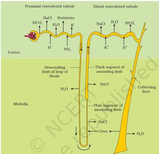
Proximal convoluted tubule (PCT)
Lined by simple cuboidal brush border epithelium, which increases the surface area for reabsorption. Nearly all the essential nutrients and 70–80% of electrolytes and water are reabsorbed here. The PCT also maintains pH and ionic balance by selective secretion of hydrogen ions and ammonia and by absorption of HCO₃⁻.
Loop of Henle
Reabsorption is minimum in its ascending limb, but the loop is significant in maintaining the high osmolarity of the medullary interstitial fluid. The descending limb is permeable to water but almost impermeable to electrolytes (concentrating the filtrate); the ascending limb is impermeable to water but allows transport of electrolytes actively or passively (diluting the filtrate).
Distal convoluted tubule (DCT)
Performs conditional reabsorption of Na⁺ and water; also reabsorbs HCO₃⁻ and selectively secretes hydrogen and potassium ions and NH₃ to maintain pH and the sodium–potassium balance of the blood.
Collecting duct
Extends from the cortex to the inner medulla. Large amounts of water can be reabsorbed here to produce concentrated urine. It allows small amounts of urea into the medullary interstitium to keep up the osmolarity, and secretes H⁺ and K⁺ selectively.
Common mistake
MCQ Questions
1 Mark Questions
- 1What is the function of the brush border in the PCT?
- 2Which segment of the nephron reabsorbs the maximum electrolytes and water?
- 3State the permeability properties of the descending and ascending limbs of the loop of Henle.
- 4Which part of the nephron allows small amounts of urea into the medullary interstitium?
- 5What does tubular secretion help the body to maintain?
2 Mark Questions
- 1Why is the ascending limb of the loop of Henle impermeable to water, and what role does this play in the medullary osmolarity?
- 2List the functions of the PCT.
- 3Explain how the DCT maintains the pH and sodium–potassium balance of the blood.
3 Mark Questions
- 1Describe the functions of the PCT, loop of Henle, DCT and collecting duct with reference to reabsorption and secretion.
- 2Discuss which substances are reabsorbed actively and which passively in different nephron segments, and how tubular secretion maintains the pH of body fluids.
Countercurrent Mechanism — Concentration of the Filtrate
Counter current mechanism
The special transport arrangement between the loop of Henle and the vasa recta: filtrate flows in opposite directions in the two limbs of the loop, and blood also flows in a counter current pattern through the two limbs of the vasa recta. This maintains an increasing osmolarity towards the inner medullary interstitium — from about 300 mOsmolL⁻¹ in the cortex to about 1200 mOsmolL⁻¹ in the inner medulla.
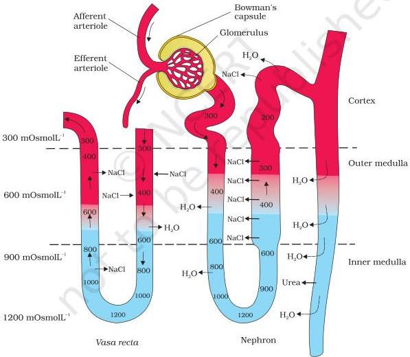
- The gradient is caused mainly by NaCl and urea. NaCl is transported by the ascending limb of the loop of Henle and exchanged with the descending limb of the vasa recta, then returned to the interstitium by the ascending portion of the vasa recta.
- Small amounts of urea enter the thin segment of the ascending limb of the loop of Henle and are transported back to the interstitium by the collecting tubule.
- The interstitial gradient allows easy passage of water from the collecting tubule, concentrating the filtrate (urine).
- Human kidneys can produce urine nearly four times more concentrated than the initial filtrate.
Exam pointer
MCQ Questions
1 Mark Questions
- 1Define the counter current mechanism.
- 2Give the osmolarity values of the kidney cortex and inner medulla.
- 3Which two substances mainly build the medullary osmotic gradient?
- 4How much more concentrated than the initial filtrate can human urine be?
2 Mark Questions
- 1Explain the role of the vasa recta as a counter current exchanger.
- 2How does the ascending limb of the loop of Henle contribute to building the medullary gradient?
- 3How does urea enter the medullary interstitium and why does this matter?
3 Mark Questions
- 1Give a brief account of the counter current mechanism and the role of the loop of Henle and vasa recta in concentrating urine.
- 2Explain why the special arrangement of the loop of Henle and the vasa recta is called a counter current mechanism, and how the interstitial gradient it builds helps concentrate the filtrate.
Regulation of Kidney Function — ADH, Renin-Angiotensin and ANF
Antidiuretic hormone (ADH) / vasopressin
Osmoreceptors are activated by changes in blood volume, body fluid volume and ionic concentration. Excessive loss of fluid activates them, stimulating the hypothalamus to release ADH from the neurohypophysis. ADH facilitates water reabsorption from the latter parts of the tubule, preventing diuresis. An increase in body fluid volume switches off the osmoreceptors and suppresses ADH release to complete the feedback.
ADH also has constrictory effects on blood vessels, increasing blood pressure; an increased blood pressure increases glomerular blood flow and hence the GFR.
Renin-Angiotensin mechanism
A fall in glomerular blood flow / blood pressure / GFR activates the JG cells to release renin, which converts angiotensinogen in the blood to angiotensin I and then to angiotensin II. Angiotensin II is a powerful vasoconstrictor that raises glomerular blood pressure and GFR; it also activates the adrenal cortex to release aldosterone, which causes reabsorption of Na⁺ and water from the distal parts of the tubule — again raising blood pressure and GFR.
Atrial natriuretic factor (ANF)
An increase in blood flow to the atria of the heart causes the release of ANF, which causes vasodilation and thereby decreases blood pressure. The ANF mechanism acts as a check on the renin-angiotensin mechanism.
MCQ Questions
1 Mark Questions
- 1What is ADH and from where is it released?
- 2What is vasopressin?
- 3Name the two hormones and one factor that regulate kidney function.
- 4What is the effect of ANF on blood pressure?
2 Mark Questions
- 1Explain the role of osmoreceptors and ADH in regulating the water balance of the body.
- 2Outline the renin-angiotensin mechanism from renin release to aldosterone action.
- 3How does ANF act as a check on the renin-angiotensin mechanism?
3 Mark Questions
- 1Describe the hormonal feedback regulation of kidney function involving the hypothalamus, the JGA and the heart.
- 2Explain how an excessive loss of fluid from the body is corrected by the ADH mechanism, and how an increase in body fluid volume completes the feedback.
Micturition and Accessory Excretory Organs
Urine formed by the nephrons is carried to the urinary bladder and stored until a voluntary signal from the CNS. Stretching of the bladder as it fills activates stretch receptors in its walls, which send signals to the CNS. The CNS sends motor messages that contract the smooth muscles of the bladder and simultaneously relax the urethral sphincter, releasing urine. This release is called micturition and the neural mechanism is the micturition reflex.
An adult human excretes on average 1–1.5 L of urine per day. Urine is a light-yellow, watery, slightly acidic fluid (pH ≈ 6.0) with a characteristic odour; on average 25–30 g of urea is excreted per day. Glycosuria (glucose) and ketonuria (ketone bodies) indicate diabetes mellitus.
Role of Other Organs in Excretion
- Lungs — remove large amounts of CO₂ (approximately 200 mL/minute) and significant quantities of water every day.
- Liver — the largest gland; secretes bile containing bilirubin, biliverdin, cholesterol, degraded steroid hormones, vitamins and drugs, which mostly pass out with digestive wastes.
- Skin — sweat glands secrete a watery fluid containing NaCl, small amounts of urea and lactic acid; sebaceous glands eliminate sterols, hydrocarbons and waxes through sebum. Small amounts of nitrogenous wastes can also be eliminated through saliva.
MCQ Questions
1 Mark Questions
- 1Define micturition and the micturition reflex.
- 2Give the average daily urine output and the average urea excretion of an adult human.
- 3Name the excretory products of the sweat glands.
- 4Which organ secretes bile containing bilirubin and biliverdin?
- 5What do glycosuria and ketonuria indicate?
2 Mark Questions
- 1Explain the neural mechanism of micturition.
- 2Describe the role of the liver and skin in excretion.
- 3Mention the characteristics of normal urine and what their analysis reveals.
3 Mark Questions
- 1Describe the role of the lungs, liver and skin in the elimination of excretory wastes.
- 2Explain micturition — how urine is stored and released through the micturition reflex.
Disorders of the Excretory System — Uremia, Hemodialysis, Renal Calculi
Uremia
Malfunctioning of the kidneys leads to accumulation of urea in the blood — a highly harmful condition that may lead to kidney failure.
Hemodialysis
Removal of urea from the blood of a uremic patient using an artificial kidney. Blood drained from a convenient artery is pumped into a dialysing unit after adding an anticoagulant (heparin). The unit contains a coiled cellophane tube surrounded by a dialysing fluid having the same composition as plasma except nitrogenous wastes. Urea and other wastes diffuse out through the porous cellophane membrane along the concentration gradient, and the cleared blood is pumped back to the body through a vein after adding anti-heparin.
- Kidney transplantation — the ultimate method for correcting acute renal failure; a functioning kidney from a donor, preferably a close relative, to minimise rejection by the host's immune system.
- Renal calculi — stone or insoluble mass of crystallised salts (e.g. oxalates) formed within the kidney.
- Glomerulonephritis — inflammation of the glomeruli of the kidney.
MCQ Questions
1 Mark Questions
- 1What is uremia?
- 2Define renal calculi.
- 3What is glomerulonephritis?
- 4Why is heparin added to blood during hemodialysis?
2 Mark Questions
- 1Describe how hemodialysis removes nitrogenous wastes using the artificial kidney.
- 2Why is a functioning kidney for transplantation preferred from a close relative?
- 3Distinguish between renal calculi and glomerulonephritis.
3 Mark Questions
- 1Explain the principle and working of hemodialysis, from blood drainage to return of cleared blood.
- 2Describe the main disorders of the excretory system — uremia, renal calculi, glomerulonephritis, and the corrective methods of hemodialysis and kidney transplantation.
NCERT Exercise Questions
From the textbook
Exercises (NCERT Q1–Q12)
- 1) Define Glomerular Filtration Rate (GFR).
- 2) Explain the autoregulatory mechanism of GFR.
- 3) Indicate whether the following statements are true or false: (a) Micturition is carried out by a reflex. (b) ADH helps in water elimination, making the urine hypotonic. (c) Protein-free fluid is filtered from blood plasma into the Bowman's capsule. (d) Henle's loop plays an important role in concentrating the urine. (e) Glucose is actively reabsorbed in the proximal convoluted tubule.
- 4) Give a brief account of the counter current mechanism.
- 5) Describe the role of liver, lungs and skin in excretion.
- 6) Explain micturition.
- 7) Match the items of column I with those of column II: Column I — (a) Ammonotelism, (b) Bowman's capsule, (c) Micturition, (d) Uricotelism, (e) ADH; Column II — (i) Birds, (ii) Water reabsorption, (iii) Bony fish, (iv) Urinary bladder, (v) Renal tubule.
- 8) What is meant by the term osmoregulation?
- 9) Terrestrial animals are generally either ureotelic or uricotelic, not ammonotelic — why?
- 10) What is the significance of juxta glomerular apparatus (JGA) in kidney function?
- 11) Name the following: (a) A chordate animal having flame cells as excretory structures. (b) Cortical portions projecting between the medullary pyramids in the human kidney. (c) A loop of capillary running parallel to the Henle's loop.
- 12) Fill in the gaps: (a) Ascending limb of Henle's loop is ______ to water whereas the descending limb is ______ to it. (b) Reabsorption of water from distal parts of the tubules is facilitated by hormone ______. (c) Dialysis fluid contains all the constituents as in plasma except ______. (d) A healthy adult human excretes, on average, ______ gm of urea per day.
Answers at a glance
- 1Q16.1 — GFR is the amount of filtrate formed by the kidneys per minute; ≈ 125 mL/min (≈ 180 L/day) in a healthy individual.
- 2Q16.2 — A fall in GFR activates the JG cells of the JGA to release renin, which stimulates glomerular blood flow and restores the GFR to normal (autoregulation).
- 3Q16.3 — (a) True; (b) False — ADH increases water reabsorption, making urine concentrated, not hypotonic; (c) True; (d) True; (e) True.
- 4Q16.4 — The loop of Henle and the vasa recta run in opposite directions (counter current). NaCl transported by the ascending limb and urea returned by the collecting tubule maintain an osmolarity gradient from 300 mOsmolL⁻¹ (cortex) to 1200 mOsmolL⁻¹ (inner medulla), allowing the kidney to produce concentrated urine.
- 5Q16.5 — Liver secretes bile with bilirubin, biliverdin, cholesterol, degraded steroid hormones, vitamins and drugs (which pass out with digestive wastes); lungs remove CO₂ (~200 mL/min) and water; skin sweat glands remove NaCl, urea and lactic acid, and sebaceous glands remove sterols, hydrocarbons and waxes.
- 6Q16.6 — Micturition is the release of urine: bladder distension stimulates stretch receptors → signals to the CNS → motor messages contract bladder smooth muscle and relax the urethral sphincter. The neural mechanism is the micturition reflex.
- 7Q16.7 — (a)–(iii) Bony fish; (b)–(v) Renal tubule; (c)–(iv) Urinary bladder; (d)–(i) Birds; (e)–(ii) Water reabsorption.
- 8Q16.8 — Osmoregulation is the maintenance of ionic balance and fluid volume (osmotic concentration) of body fluids by excretory structures such as protonephridia (flame cells) and nephridia.
- 9Q16.9 — Ammonia is highly toxic and needs large volumes of water for elimination; terrestrial animals conserve water by converting ammonia to the less toxic urea (ureotelic) or almost insoluble uric acid (uricotelic), which need far less water.
- 10Q16.10 — The JGA autoregulates GFR: on a fall in GFR, its JG cells release renin, which restores glomerular blood flow and hence normal GFR.
- 11Q16.11 — (a) Amphioxus (a cephalochordate); (b) Columns of Bertini (renal columns); (c) Vasa recta.
- 12Q16.12 — (a) impermeable, permeable; (b) ADH (vasopressin); (c) nitrogenous wastes; (d) 25–30 gm.
CBSE Pattern Questions
How CBSE asks this chapter
MCQ Questions
1 Mark Questions
- 1Define GFR and give its value.
- 2Give the osmolarity range from the kidney cortex to the inner medulla.
- 3Name the hormone that prevents diuresis.
- 4What is renal calculi?
- 5Define micturition.
2 Mark Questions
- 1Why are reptiles and birds uricotelic?
- 2Explain tubular secretion and its significance.
- 3State the significance of the juxta glomerular apparatus.
- 4Distinguish between cortical and juxta medullary nephrons.
3 Mark Questions
- 1Explain the counter current mechanism of urine concentration.
- 2Describe how ADH regulates the water balance of the body.
- 3Describe the functions of the PCT and the loop of Henle.
5 Mark Questions
- 1(a) Describe the three processes of urine formation — filtration, reabsorption and secretion — with a note on the JGA. (b) Explain how ADH and aldosterone regulate water and ionic balance.
- 2(a) Give a labelled account of a nephron. (b) Describe the principle and working of hemodialysis. (c) Why is the ureotelic mode of excretion advantageous to mammals?
PDF: Assignments & Tests
Printable study material
Free PDF Downloads & Assignments
Practice path: complete Assignment 1 first (topic-by-topic), then Assignment 2 (higher difficulty, with integrated problems), then attempt the two timed CBSE-pattern tests.
Walks every topic of the chapter in order — modes of excretion, nephron structure, urine formation, tubule functions, countercurrent mechanism, regulation, micturition and disorders.
Higher-difficulty second set with integration of multi-topic problems — concentration gradient calculations, hormone feedback pathways and diagram-based questions on the nephron and countercurrent mechanism.
Full board-pattern test: 1M MCQs/VSA, 2M, 3M, 5M long answers → totals 40 marks; time 90 minutes; answer key included.
Second full paper with different questions, same 40-mark / 90-minute blueprint — urine formation, countercurrent mechanism, hormonal regulation and disorders, with answer key.
Quick Revision
Key formulas at a glance
Memorise these equations — direct application numericals and derivations in CBSE & JEE frequently hinge on these.
Glomerular filtration rate
amount of filtrate formed per minute by the kidneys.
Blood filtered per minute
ultrafiltered at the glomerulus.
Urine volume
Reabsorption percentage
Medullary osmolarity gradient
built by NaCl and urea.
Urine pH
Urea excreted per day
CO₂ removed by lungs
Exam Strategy
How this chapter is asked
High-yield question patterns observed across CBSE boards, JEE Main & Advanced, and NEET.
- About 1200 mL of blood is filtered per minute — roughly 1/5 of the blood pumped out by each ventricle.
- GFR ≈ 125 mL/min = 180 L/day; only 1–1.5 L is voided as urine, so >99% of the filtrate is reabsorbed.
- The PCT reabsorbs nearly all essential nutrients and 70–80% of electrolytes and water — the main site of reabsorption.
- Ultrafiltration: podocytes leave filtration slits, so the filtrate is essentially protein-free plasma.
- JGA autoregulates GFR — a fall in GFR triggers renin release, which restores glomerular blood flow.
- Countercurrent mechanism builds a medullary osmolarity gradient of 300 mOsmolL⁻¹ (cortex) → 1200 mOsmolL⁻¹ (inner medulla), produced mainly by NaCl and urea.
- ADH (vasopressin) from the neurohypophysis facilitates water reabsorption from the latter parts of the tubule, preventing diuresis.
- RAAS: renin → angiotensin I → angiotensin II (vasoconstrictor) → aldosterone → Na⁺ and water reabsorption; ANF opposes this by vasodilation.
- Bony fishes are ammonotelic; mammals and marine fishes are mostly ureotelic; reptiles, birds, insects and land snails are uricotelic.
- An adult excretes about 25–30 g of urea daily; glycosuria and ketonuria point to diabetes mellitus.
- Hemodialysis uses a dialysing fluid identical to plasma except nitrogenous wastes, over a porous cellophane membrane; heparin is added as an anticoagulant.
Practice Numericals
Step-by-step worked examples
Standard exam-level problems solved with clear conceptual breakdowns.
Percentage of filtrate reabsorbed
Step-by-Step Solution
- 1Total filtrate per day = 125 mL/min × 60 min × 24 h = 180,000 mL = 180 L.
- 2Urine produced = 1.5 L, so reabsorbed = 180 − 1.5 = 178.5 L.
- 3Percentage reabsorbed = (178.5 / 180) × 100 ≈ 99.2%.
- 4This confirms that over 99% of the filtrate is reabsorbed, mainly by the PCT.
Final Answer
≈ 99.2%
Effect of water load on urine concentration
Step-by-Step Solution
- 1When water intake is high, blood osmolarity falls, osmoreceptors are suppressed, and the hypothalamus reduces ADH release from the neurohypophysis.
- 2With low ADH, the latter parts of the tubule reabsorb little water, so a large volume of dilute urine is produced.
- 3During dehydration, blood osmolarity rises, osmoreceptors activate, ADH release increases, and more water is reabsorbed — producing a small volume of concentrated urine.
- 4This is the ADH feedback loop that prevents diuresis and conserves body water.
Why the medullary gradient concentrates urine
Step-by-Step Solution
- 1The gradient is built by the countercurrent arrangement of the loop of Henle and vasa recta, accumulating NaCl and urea in the medullary interstitium.
- 2As filtrate (and later urine) passes down the collecting duct through this high-osmolarity interstitium, water is drawn out by osmosis.
- 3The nearer the medulla, the greater the osmotic pull, so the final urine becomes highly concentrated.
- 4Because of this mechanism human kidneys produce urine nearly four times more concentrated than the initial filtrate.
Final Answer
≈ 4× the filtrate
FAQ
Frequently asked questions
What are the three processes of urine formation?
The three processes are: (1) glomerular filtration — ultrafiltration of blood at the glomerulus (GFR ≈ 125 mL/min); (2) tubular reabsorption — recovery of essential nutrients, electrolytes and water, mainly in the PCT (>99% of the filtrate); and (3) tubular secretion — transfer of H⁺, K⁺ and ammonia from blood into the filtrate to maintain pH and ionic balance.
What is the role of ADH (vasopressin) in kidney function?
When body fluid volume or ionic concentration changes, osmoreceptors activate the hypothalamus to release ADH from the neurohypophysis. ADH facilitates water reabsorption from the latter parts of the tubule, preventing diuresis and producing concentrated urine. An increase in body fluid volume switches off the osmoreceptors and suppresses ADH release to complete the feedback.
How does the countercurrent mechanism concentrate urine?
The loop of Henle and the vasa recta run in opposite directions, forming a counter current. NaCl transported by the ascending limb and urea returned by the collecting tubule maintain an osmolarity gradient from 300 mOsmolL⁻¹ (cortex) to 1200 mOsmolL⁻¹ (inner medulla). Water is drawn out of the collecting duct by this interstitial gradient, so the kidney produces urine nearly four times more concentrated than the filtrate.
Why are terrestrial animals ureotelic or uricotelic and not ammonotelic?
Ammonia is the most toxic nitrogenous waste and needs large volumes of water for elimination. Terrestrial animals conserve water, so they convert ammonia into the less toxic urea (mammals — ureotelic) or into almost insoluble uric acid excreted as a pellet (reptiles, birds — uricotelic). Uric acid needs the least water of all.
What is hemodialysis and when is it needed?
Hemodialysis removes urea accumulated in the blood (uremia) using an artificial kidney. Blood is drained from an artery, mixed with heparin, and passed through a coiled cellophane tube immersed in a dialysing fluid identical to plasma except nitrogenous wastes; wastes diffuse out and the cleared blood is returned with anti-heparin. It is needed in kidney failure, and kidney transplantation is the ultimate corrective method.
What is the significance of the juxta glomerular apparatus (JGA)?
The JGA is a region formed by modifications in the DCT and afferent arteriole at their contact. A fall in GFR activates its JG cells to release renin, which stimulates glomerular blood flow and brings the GFR back to normal — the autoregulatory mechanism of the kidney.
Which structures perform excretion in invertebrates?
Protonephridia (flame cells) in Platyhelminthes, rotifers, some annelids and Amphioxus; nephridia in earthworms; malpighian tubules in insects; and antennal (green) glands in crustaceans like prawns. They remove nitrogenous wastes and regulate ionic/fluid balance (osmoregulation).
What do lungs, liver and skin excrete?
Lungs remove CO₂ (≈ 200 mL/min) and water; the liver secretes bile carrying bilirubin, biliverdin, cholesterol, degraded steroid hormones, vitamins and drugs; skin sweat contains NaCl, urea and lactic acid, and sebaceous glands eliminate sterols, hydrocarbons and waxes.
Next Chapters
Continue learning
Same chapter
Read alongside
Interactive Quiz
Chapter MCQ practice test
Instant scoring with complete solutions — test your mastery in under 15 minutes.
Chapter MCQ Mock Test
Evaluate how well you have retained the concepts, formulas, and reaction mechanisms from this chapter. Questions adhere strictly to latest CBSE, JEE & NEET trends.
Master this chapter with expert live guidance
Self-study notes lay the ground, but conceptual doubts clear fastest in an interactive classroom. Narayan Gurukul Academy (ClassApna) conducts small-batch CBSE, JEE & NEET coaching with daily doubt solving and rigorous mock tests.
Small batches · 1-on-1 personal mentorship · Live online & offline centre